

Break through the bottleneck! 3D printing + stem cell technology to help nerve regeneration
Release time:
2025-08-29
Spinal cord injury is the "incurable injury" in the global medical community. On August 23, the University of Minnesota team published a major study in "Advanced Medical Materials", which opened a light for this dilemma: using 3D printing technology to build a "smart scaffold" and combined with the neural progenitor cells cultivated by stem cells, it successfully set up a neural pathway in the body of rats with completely broken spinal cord, so that they can restore their walking ability after 12 weeks.
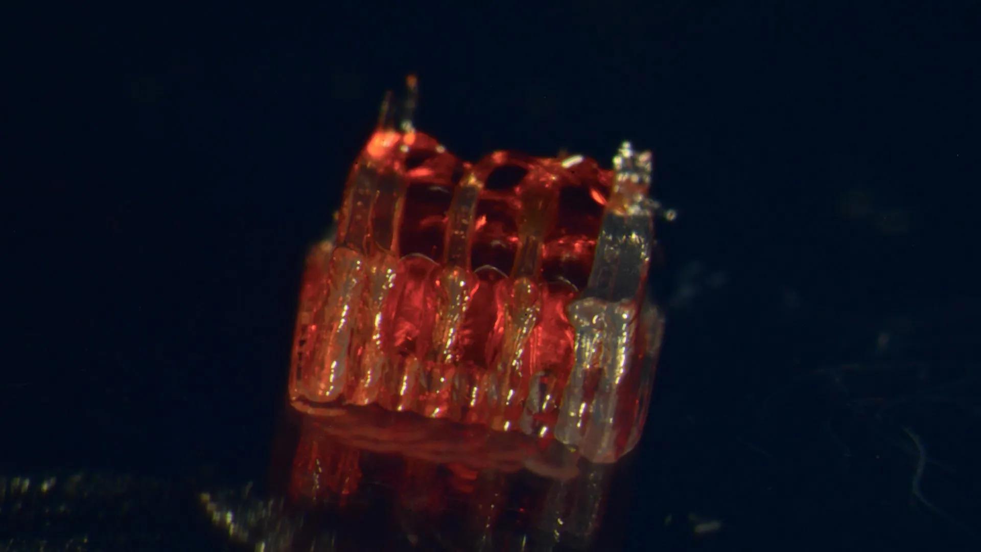
Spinal cord injury: The century-old dilemma of "incurable injuries" in the medical community
The spinal cord is the "information highway" connecting the brain and the body. Once it is completely broken due to a car accident, fall, or other accidents, the nerve signals will be completely interrupted, resulting in paralysis. What’s even more difficult is that central nervous cells almost lose their natural regeneration ability. Traditional treatment methods (such as rehabilitation training, drug intervention) can only relieve symptoms and cannot repair the broken neural circuit.
In the past, the treatment methods of direct injection of stem cells often resulted in disorderly proliferation of cells and made it difficult to form functional neural connections due to the lack of effective guidance for cell growth. This core problem has long restricted the progress of spinal cord injury treatment, until the intersection of 3D printing technology and stem cell biology provided a new path for breaking the deadlock.
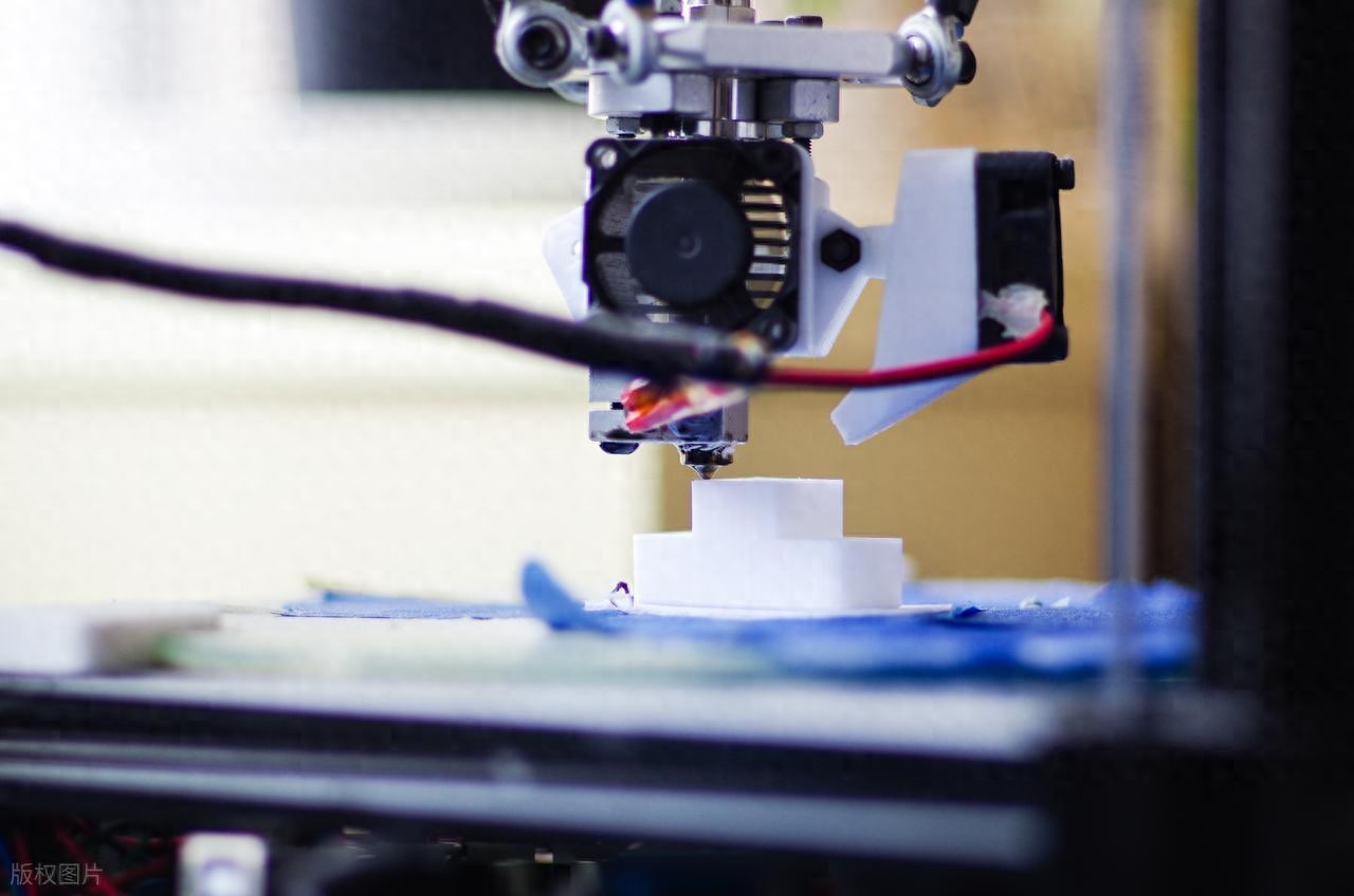
3D printing + stem cells: "Smart Scaffold" builds a high-speed road for nerve regeneration”
In response to this challenge, the research team of the Department of Mechanical Engineering of the University of Minnesota deeply integrates the "precision control" of engineering with the "regenerative potential" of biology to create the revolutionary tool of "organoid scaffolding".
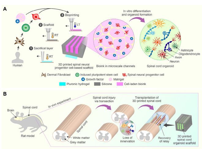
The research team first 3D printed the microchannel scaffold using biocompatible materials - these microchannels with a diameter of only 200 microns (about the thickness of a hair) to simulate the microenvironment of normal spinal cord tissue, just like laying a "highway with guardrail" for nerve fibers to ensure that they grow in the target direction. Subsequently, the team injected the "spinal cord nerve progenitor cells" differentiated from human adult stem cells into the scaffold. These "nerve seeds" proliferate and differentiate under the guidance of the scaffold, and after 40 days, a highly ordered neural network - the "mini spinal cord" organoid was "grown" in vitro.
The study pointed out that this "in vitro prefabrication + in vivo transplantation" model effectively avoids the disorder of traditional cell transplantation through physical guidance of the scaffold structure, and provides key technical support for the precise reconstruction of neural circuits.
Rat experiment succeeded: After 12 weeks of paralysis, they stood up again
To verify the effectiveness of the technology, the team selected rats with complete spinal cord fracture (the most severe injury model) for experiments. After transplanting the "mini spinal cord" stent to the injured area, the miracle gradually appeared:
Motor function recovery: 12 weeks after surgery, the BBB motor function score (a professional scale for estimating hindlimb motor ability) in the transplanted rats reached 8.4 points, while the scores of the untreated group and the control group with only empty stent implantation were only 2.25 and 3.6 points. These once completely paralyzed rats can not only support their weight, but also coordinate their limbs to walk.
Neural pathway reconstruction: Electrophysiological tests show that the electrical signals emitted by the brain of rats in the treatment group can be transmitted to the lower limb muscles through new neural circuits, confirming that the "information highway" has been successfully penetrated.
Cell fusion verification: Microscopy observed that human-derived nerve cells not only survive, but also actively extend synapses, "shake hands" with rats' spinal cord nerve endings, forming a stable new connection.
The researchers said this result marks a key step in the field of spinal cord injury treatment. At present, the team is advancing technology optimization: developing degradable scaffolds (avoiding foreign body residue), exploring more complex neural cell combinations (reconstructing sensory functions), and accelerating clinical transformation through multidisciplinary collaboration.
Although clinical applications from rats to humans still need to overcome challenges such as large-scale production and safety assessment, this study undoubtedly illuminates the light of hope for 300,000 spinal cord injury patients around the world - the once "irreversible" paralysis is being re-written by the power of science and technology.
Latest developments

The debut and discourse of the YinFeng Life Continuation Project not only served as an authoritative demonstration of its own ethical governance framework but also contributed practical wisdom toward establishing industry norms.

Yinfeng Foundation Honored as "2025 Public Welfare Donor" by Jinan Red Cross
Looking ahead, Yinfeng Foundation will use this recognition as an opportunity to further deepen its strategic collaboration with the Jinan Red Cross, expand cooperation in areas such as life health and emergency response, and continue to empower efforts to accelerate the construction of a "new, strong, excellent, rich, beautiful, and high-quality" modern socialist strong capital city, contributing even more Yinfeng strength to this endeavor.

The team successfully developed the CryoSIM platform, an intelligent microfluidics and deep learning-integrated system. This platform deeply integrates core technologies of deep learning and microfluidics to enable high-throughput, high-precision automated analysis of oocyte membrane permeability. It provides a novel technological tool for optimizing and advancing the clinical translation of oocyte cryopreservation techniques. Additionally, it offers an innovative practical paradigm for the application of artificial intelligence in low-temperature biomedicine and reproductive medicine.

Public Welfare Partnership: A Special Letter from the Jinan Red Cross
On the afternoon of January 30, 2026, the Jinan Red Cross presented a letter of special significance to the Shandong Yinfeng Life Science Public Welfare Foundation (hereinafter referred to as the Yinfeng Foundation).

World’s First Achievement Highlights Brand Leadership
In the future, Yinfeng Life Science Research Institute will continue to uphold its mission of "Dedicated to Medical Technology, Safeguarding Human Health." It will empower brand building with more original and pioneering scientific and technological achievements, contributing wisdom and strength to Shandong's goal of building a national regional innovation hub and promoting Chinese brands on the global stage.

Global First Ovarian Tissue Dual Activation Technology Debuts at 2025 Jinan Achievements Conference
Currently, the ovarian tissue dual activation technology has been successfully applied in clinical practice at Beijing University of Chinese Medicine Shenzhen (Longgang) Hospital, having treated over 400 patients with a treatment success rate of 70%. Over the next three years, Shandong Yinfeng Life Science Research Institute plans to use Jinan as a center to gradually expand the transformation and application of this technological achievement nationwide.
Yinfeng Life Continuation Plan
Address: Yinfeng Biotechnology Park, 1758 Gangyuan 6th Road, High-tech Zone, Jinan City, Shandong Province
Zip Code: 250102
Telephone: 0086-0531-88897351
Mobile: 0086-19953155515

Mobile website

Official WeChat Public Number
Links:Red Cross Society of China丨Shandong Red Cross Society丨Shandong Provincial Health Commission | Yinfeng Group| Yinfeng Biological Group
All rights reserved©Yinfeng Life Continuation Powered by www.300.cn SEO